

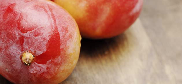
The Australian mango fruit grower’s market is a substantial industry sector that is anticipated to grow by 20% in 2014. The ripening, transportation, and saleable life-span of mangoes depends on keeping the fruit free from pests and disease.
 |
| The use of irradiation is emerging as a viable, chemical-free alternative to traditional pesticides. |
Worldwide, the use of irradiation is emerging as a viable, chemical-free alternative to traditional pesticides.
This research builds upon our knowledge in understanding the physiological responses of three new Australian mango fruit hybrids to varying doses of gamma irradiation following harvest. Of particular interest is the question of whether irradiation may degrade the fruit, such as cause damage to lenticels (small pores) on the fruit’s outer skin.
The study illustrates how ANSTO’s expertise makes a crucial contribution to the development of Australia’s national food security by providing new insight into the viability of irradiation to achieve safe phytosanitary (pest and disease) protocols for the agriculture industry.
Using magnetic resonance imaging for studying mangoes
Magnetic resonance imaging (MRI) offers a means of non-destructively examining fruit for internal and surface defects and has been used here to monitor changes in the water status of tissues of fruits and flowers [1-4]. MRI is distinct from most other imaging modalities in being able to provide multiple methods of contrast formation dependent on biophysical properties across a wide range of spatial scales.
For example, T2-weighted MRI is dependent on the interactions of water molecules with their mmediate molecular environment. In fruit, interactions with macromolecules including carbohydrates and proteins predominate. Alternatively, diffusion-weighted MRI produces images in which contrast depends on the displacement of water molecules over a specific time period (typically 10-100 ms) in a specific direction. As water movement is restricted by intracellular structures and cell walls DWI can be used as a probe of tissue pathology and degradation.
The mangoes investigated
In this trial, we investigated the effect of gamma irradiation on internal browning and bruising of a small number of NMBP1243 mangoes, see Fig.1. Eight mangoes were gamma-irradiated with doses of 0, 200 or 800 Gy using ANSTO’s cobalt-60 irradiation facility, at a dose rate of 10.1 Gy.min-1. Fricke (ferrous ammonium sulphate) dosimeters were used to monitor the irradiation doses.
Tissue changes were assessed by MRI immediately after (0 hr) and 43 hrs after irradiation, see Fig. 2. MR images of mango 2 (800 Gy, indicated with red square in photo) are shown. The images illustrate internal mango flesh (mesocarp) browning developed over two days but not visible as defect or discolouration on the skin (epicarp). There was no evidence of tissue damage relative to the two unirradiated samples.
In diffusion-weighted imaging, signal strength (image contrast) is dependent on the physical restriction of water movement especially by cell walls and organelles. Fig. 3 demonstrates an extensive region of increased water diffusivity (orange pixels).
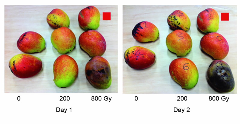 |
| The mangoes investigated: NMBP143 gamma-irradiated with doses of either 0,200 or 800 Gy, on the right after two days. |
Conclusion
Both T2 and diffusion-weighted MRI clearly illustrated mango tissue changes associated with internal browning, some of which were not visible on the skin.
There was no evidence of changes in tissue properties associated with irradiation. MRI may also be valuable in investigations of radiation damage to lenticels. In this case samples of mango skin would be imaged at very high spatial resolution in an 11.5 or 16 Tesla microimaging system.
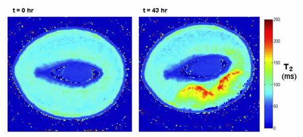 |
| Qualitative imaging (T2 weighted) of the development of internal mango browning (imaging was performed on a GE Syngo 3T MRI system with 8-channel transmit/receive knee coil). Mesocarp (internal) browning (yellow and red area), evident as a 50-100% increase (in T2), is consistent with sugar hydrolysis and decreased local sugar concentration. In this example the absence of change in extent of the dark blue (low-T2 ) ‘halo’ around the seed illustrates the absence of significant ripening progression over the 43 hour measurement interval (consistent with absence of change in skin colour of this specimen. |
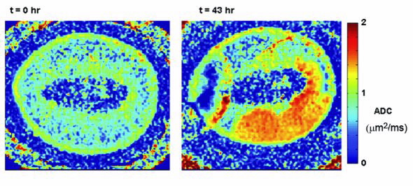 |
| Apparent diffusion coefficient (ADC) image of the development of mango browning (cf. Fig 2). Mesocarp (internal) browning is evident as 50-100% increase in ADC consistent with the breakdown of cell walls over an extended volume of tissue. (Concentric rings are a parallel image artefact which can be eliminated by use of an alternative imaging protocol. |
Authors
Roger Bourne2, Connie Banos1, Justin Davies1,2 Richard Banati1,2, Robert Henriod3
1ANSTO, 2University of Sydney, 3Queensland Department of Employment, Economic Development and Innovation
References
- Joyce, D.C., et al., H-1-Nuclear magnetic resonance imaging of ripening ‘Kensington Pride’ mango fruit. Functional Plant Biology, 2002. 29(7): p. 873-879.
- Clark, C.J. and MacFall, J.S., Quantitative magnetic resonance imaging of ‘Fuyu’ persimmon fruit during development and ripening. Magnetic Resonance Imaging, 2003. 21(6): p. 679-685.
- Musse, M., et al., Monitoring the postharvest ripening of tomato fruit using quantitative MRI and NMR relaxometry. Postharvest Biology and Technology, 2009. 53(1-2): p. 22-35.
- Yooyongwech, S., et al., Changes in aquaporin gene expression and magnetic resonance imaging of water status in peach tree flower buds during dormancy. Physiologia Plantarum, 2008. 134(3): p. 522-533.
Published: 13/09/2012