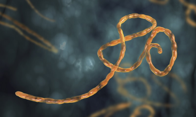
The International Atomic Energy Agency (IAEA) will provide specialised diagnostic equipment to help Sierra Leone in its efforts to combat an ongoing Ebola Virus Disease (EVD) outbreak.
 |
| Ebola Virus Digital Illustration |
The support is in line with a UN Security Council appeal and responds to a request from Sierra Leone. The IAEA assistance will supplement the country's ability to diagnose EVD quickly using a diagnostic technology known as Reverse Transcriptase Polymerase Chain Reaction (RT-PCR).
The assistance, expected to be delivered in coming weeks, initiates broader IAEA support to African Member States to strengthen their technological abilities to detect diseases transmitted from animals to humans - zoonotic diseases.
The IAEA and the Food and Agricultural Organization of the United Nations have been at the forefront of developing RT-PCR, a nuclear-derived technology which allows EVD to be detected within a few hours.
Background information about nuclear techniques used for Ebola Virus diagnostics
At ANSTO, researchers routinely use a combination of highly sensitive bioanalytical and nuclear techniques to address important medical issues.
The technique reported by the IAEA is RT-PCR or ‘reverse transcriptase – polymerase chain reaction’. In this technique, minute amounts of genetic material (RNA) from a virus, such as Ebola, is re-written (the technical term is ‘reverse transcribed’) into DNA. From this DNA, copies as well as copies of copies are made by a ‘polymerase chain reaction’ leading to a massive amplification of what initially was only a small amount of genetic material.
During this process of amplification, one of the building blocks of the genetic material needed to make the copies, the nucleotide deoxycytidine triphosphate (dCTP), is added. In order to make the incorporated nucleotide detectable, it is labelled with phosphorus-32, a radioisotope with a short half-life of 14.29 days.
Alternatively, a short, phosphorus-32–labelled sequence of nucleotides that specifically matches one of the sequences found in the virus is added later to the amplified DNA copies after they have been blotted onto filter paper, a method called a dot blot hybridization. The darkened spots show which of the tested samples, for example from blood, contain the virus.
Alternatively, a short, phosphorus-32–labelled sequence of nucleotides that specifically matches one of the sequences found in the virus is added later to the amplified DNA copies after they have been blotted onto filter paper, a method called a dot blot hybridization. The darkened spots show which of the tested samples, for example from blood, contain the virus.
In either approach, it is the phosphorus-32 which provides the measurable signal. It is detected by exposing the gel or filter with the amplified copies to a photographic film or plate, a process called ‘autoradiography’. Based on the initial work using radioisotopes, the technique has been refined and it is now possible to replace phosphorus-32 with fluorescent labels.
Professor Richard Banati (NST – Strategic Research) says: “Radioisotopes are strategically important for technology development in medicine. In certain situations where cost, speed and volume in the absence of expensive laboratory equipment and reagents are an issue, radioisotope-labelled probes continue to be developed for large-scale screening of blood samples and for surveillance purposes.”
At ANSTO, RT-PCR and autoradiography are used for a range of important bioanalytical questions. They are continuously refined and developed for new targets and applications.
“We have been amplifying the genetic material from microbes to human patients and even 3000 year old sheep from an archaeological dig.
The combination of RT-PCR and radioisotopes is fascinating: one technique (RT-PCR) uses an enzyme to copy and massively amplify genetic information; the other uses radioisotopes to detect the smallest amount of matter.
Together, this is one of the most sensitive measurement techniques in biology and medicine.”


