

 |
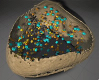
An example of the power of visual proteomics (the study of proteins). Photo credit:Max Planck Institute of biochemistry |
Advances in cryoelectron microscopy and non-invasive high resolution imaging methods are providing fundamental insights into cellular organisation and structure in their native environment that will benefit the life science and medicine, according to biophysicist and biochemist Professor Wolfgang Baumeister of the Max Plank Institute in Munich, Germany.
At present, cryoelectron tomography is the only technique that can be used for deeply buried folded proteins or coiled molecules that may be fragile or transient. The approach, which involves snap freezing samples in vitreous ice, is expected to be used to model the progression of diseases such as Parkinson’s and Alzheimer’s and in designing therapeutic drugs.
Prof Baumeister shared some dynamic tomographic slices and 3D visualisations of large biological molecules in intact cells under close-to-life conditions that provide information about the architecture and molecular interactions at the atomic scale.
“They are bridging the gap between molecular and cellular biology,” said Prof Baumeister , who pioneered the development of cryoelectron tomography.
He was at ANSTO on 6 February for the first time to deliver a Distinguished Lecture as part of a series that features leading thinkers from science and technology.
He was at ANSTO on 6 February for the first time to deliver a Distinguished Lecture as part of a series that features leading thinkers from science and technology.
Monash University, an ANSTO partner, has recently purchased a cryoelectron microscope that will be available to Australian researchers. This multi-modal semi-automated technology can be used for 2D and 3D characterisation of biological samples with electron crystallography, single-particle analysis, cryoelectron microscopy, and dual-axis cellular tomography.
“The exceptional quality and definition of sub-cellular and protein structures using complementary imaging technologies, opens a window with a breathtaking view on biomolecular processes, which means Prof Baumeister continues to drive the technical development as well as the ever more powerful computational data analysis and data integration,” said Biomedical Scientist Richard Banati, another pioneer in nuclear imaging for the life sciences.
The three types of microscopy have emerged that are helping in mapping the molecular landscape inside cells are: electron crystallography, single-particle cryoelectron microscopy and cryoelectron tomography. Together they provide an approach to imaging biological structures from molecules to cells.
Prior to the development of cryoelectron tomography, the imaging of one-of-a-kind heterogeneous entities had been the most significant obstacle to visualising subcellular detail, explained Prof Baumeister.
Prof Baumeister combined techniques in seminal research characterising the 26S proteasome, a complex molecular machine built from 31 subunits, which regulates protein degradation within the cell and is implicated in a number of diseases.
In 2012, Baumeister et al published a paper in the Proceedings of the National Academy of Sciences describing an integrative approach based on data from cryoelectron microscopy, X-ray crystallography, residue-specific chemical cross-linking, and several proteomics techniques.
His most recent paper was published in Science in January, provides more detailed information and 3D images of the capped regulatory subunits of the 26S proteasome in intact neurons.
The rapid freezing of the sample in amorphous ice below 160⁰C to preserve structures, 3D tomographic reconstruction and the use of a Volta phase plate that enables in-focus phase contrast are providing the best possible picture of cellular environments including structures deeply buried within a complex structure, such as the proteasome.
“There is an enormous richness of information available with this cryoelectron tomography. It is a game changing technology,” said Prof Baumeister.
Focused ion beam milling using gallium to create pressure-free samples thin enough for tomography and dual tilting of the sample improves information content significantly.
“Improvements in computational power have also reduced what used to take three and a half years to complete to about eight hours of data collection.”