

Radiation transport simulations were compared with experimental measurements obtained at the High Energy Reference Field facility at CERN (European Organization for Nuclear Research, Geneva, Switzerland) to provide an improved understanding of this complex mixed radiation-field used in the calibration and testing of dosimetry instrumentation.
 |
In addition, a performance evaluation of a recently developed solid-state microdosimeter was completed. Both aspects of this study contribute to radiation-biology research and improved radiation-protection practises for commercial aviation, space sciences and high-energy physics.
Dale Prokopovich1, Mark Reinhard1, Anatoly Rosenfeld2, Iwan Cornelius2 and Graeme Taylor3 1ANSTO, 2University of Wollongong, Australia, 3National Physical Laboratory, UK |
The CERN - EU (European Union) High Energy Reference Field facility
In order to improve radiation protection for air crew and astronauts we combine radiation transport simulations and experimental measurements to further the understanding of the complex mixed radiation-fieldattheHighEnergyReferenceField (CERF) facility [1]. An improved understanding of the complex radiation field allows greater accuracy of dosimetry equipment calibrated at the facility. In addition, we tested a new solid-state microdosimeter device developed in Australia for performing radiation-protection measurements in unknown mixed radiation fields.
The CERF facility generates a radiation field with the characteristics of a cosmic-ray generated background found in the upper atmosphere.
A neutron-dominated field of similar spectral characteristics is produced via the interaction of a high energy 120 GeV/c mixed hadron beam with a thick copper target and subsequent transport of the secondary particle production through thick concrete walls to simulate the cascade of secondary particles through the upper atmosphere.
CERF is predominantly used in the calibration of radiation-dosimetry instrumentation for applications in aviation, space and highenergy physics and for the evaluation of new dosimetry instrumentation and measurement techniques.
The complex environment of CERF was simulated,usingtheMonteCarlo-basedGEANT4 toolkit [1], to recreate the particle-energy fluence of the radiation field at different positions of interest within the facility. Figure 1a-d displays the various components of the particle-energy fluence from the mixed-particle radiation field atthe CERF facility irradiation position CS1, consisting of high-energy particles including neutron, proton, muon, pion, electron, positron and gamma-ray components.
The mixedradiation field is dominated by a broad energy spectrum of neutrons and similarly gamma radiation, as shown in Figure 1a. Knowledge of these two components is well understood. In particular the dominant neutron component compared well with data obtained from simulations [2] using the FLUKA Monte Carlo transport-code, and from published experimental Bonner sphere measurements [3].
Of considerable interest is the non-negligible presence of a charged-particle component not previously considered relevant to the calculation of dosimetry calibrations factors employed at the facility. The current simulations revealed a significant fluence component of electrons, positrons (Figure 1b) and protons (Figure 1a) in addition to other charged particles, pions (Figure 1d) and muons (Figure 1c), at lesser fluence.
Prior to these simulations the charged-particle component was considered negligible in terms of the contribution to the measurable dose rate. These results suggest the importance of including the charged-particle component into the facility’s dosimetry calibration factors.
Microdosimeters
Microdosimetry provides a means to obtain a radiological hazard assessment of complex mixed radiation-fields. Conventionally, this is performed using a Tissue Equivalent Proportional Counter (TEPC) filled with a tissue equivalent gas at low pressure (0.4 - 1 kPa).
As an alternative we developed a solid-state detector using a silicon-on-insulator (SOI) microdosimeter with the advantages of low weight, low voltage of operation, low power consumption and low maintenance, while being able to monitor a wide range of particles and energies [4].
The SOI Microdosimeter device possesses an array of human cell sized (30 x30 x 10µm3) sensitive detection volumes used to measure the spectrum of lineal energy deposit events when exposed to the field of interest.
Lineal energy is the ratio of the energy deposited in a volume of tissue to the average chord length of that volume (measured in units of keVµm-1). Using the regional microdosimetry approach [5] the lineal energy deposition spectrum, along with radiobiological data, is used to estimate the radiological hazard.
Comparison measurements using both a commercial TEPC (HAWK) and the SOI Microdosimeter were undertaken at identical positions within the CERF field. In the case of the SOI Microdosimeter, data were acquired using both a bare device and additionally with an over layer of low-density polyethylene to produce recoil protons from the neutron component of the field.
The SOI Microdosimeter spectra were adjusted using a geometric tissue equivalence scaling factor for silicon to tissue dose conversion of z=0.63 [6] and a correction for charge collection efficiency of 80% at the 10 V operating bias employed [7].
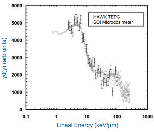 |
| Figure 2: Comparison of the lineal energy spectra as obtained with the SOI Microdosimeter and commercial HAWK TEPC. |
Suitability of a silicon-on-insulator microdosimeter
Figure 2 shows a good agreement between the TEPC and the SOI Microdosimeter measurements over the full range of lineal energies. This indicates that the SOI Microdosimeter is suitable for use in the radiation environment encountered at the CERF facility in comparison with conventional TEPC microdosimetry techniques.
The SOI Microdosimeter response to the CERF radiation field was also simulated (using GEANT4) for the various field components. The experimental measurement and the simulated response of the combined radiation field components compare well for the range of lineal energies covered, as shown in Figure 3.
Using our simulations we can show that the majority of the contribution for the mid-range of lineal energies comes from the charged-particle component of the field. By comparing the results for the SOI Microdosimeter, the TEPC and the GEANT4 simulation, it can be demonstrated that the charged-particle component contributes significantly to the microdosimetric spectrum. The contribution of this charged-particle component was not recognised in previous microdosimetry measurements.
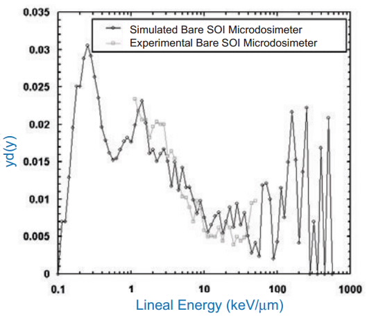 |
| Figure 3: Comparison of the simulated and experimentally measured bare SOI Microdosimeter lineal energy spectra. |
Conclusions
GEANT4 simulations of CERF have revealed the non-negligible presence of a charged-particle component of the radiation field. The accurate calibration and testing of dosimetry equipment undertaken at the facility requires that the dose contributions of the charged-particle component are properly taken into account.
Testing of a new solid-state microdosimeter device demonstrated comparable performance to existing TEPC instruments. The SOI Microdosimeter is a small, low weight, battery operable device which does not require a gas supply and can provide a true on-line measurement of the microdosimetric properties of any arbitrary mixed radiation field.
It is highly suited as a portable instrument for radiation protection applications in high-energy mixed radiation fields for aviation dosimetry and related applications in space sciences and high-energy physics.
A new generation of SOI Microdosimeter is currently under development (Australian Research Council grant between University of Wollongong, University of New South Wales and ANSTO).
This new generation of SOI Microdosimeter will provide the capability of online personal dosimetry in radiation-protection applications such as nuclear reactors, high energy accelerators as well as aviation and space applications. As soon as the development is completed, the new devices will be tested at the CERF facility.
References
- Agostinelli et al., Nuclear Instruments and Methods in Physics Research A, 506, (2003) 250-303.
- MitaroffAand Silari M, Radiation Protection Dosimetry, 102(1), (2002) 7–22.
- IAEA, Technical Reports Series, No. 403, (2001) ISBN 92–0–102201–8.
- RosenfeldA. B and Bradley P. D, Radiation Protection Dosimetry, 85(1-4), (1999) 385-388.
- Rossi H.H and Zaider M, Springer (1996) ISBN 3-54058541-9.
- Bradley P. D and RosenfeldA. B, Medical. Physics, 25, (1998) 2220–2225.
- Cornelius I, RosenfeldA, Siegele R, and Cohen D, IEEE Transactions on Nuclear Science, 50, (2003) 2373–2379.
Published: 29/09/2014