New perspectives on paleontological samples are being opened up by non-invasive neutron radiography and tomography on ANSTO’s “Dingo” instrument, which reveal surprising compositional and textual information that is not available from other techniques.
The information is producing startling findings about the evolutionary timeline, and the behaviour of creatures that lived up 1.5 billion years ago.
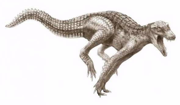 |
| Fossils from a Cretaceous crocodylomorph, an ancestor or close relative to modern-day crocodiles, have been analysed and imaged on Dingo Image © : Todd Marshall/National Geographic |
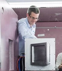
“Using neutrons for computed tomography (CT) scans can actually work better in some cases than using X-rays to determine features of fossils, bones and tissues that have been turned to rock over millions of years, and which are themselves encased in rock,” said Joseph Bevitt (below right), ANSTO Science Coordinator, manager of the neutron instruments User Office and Dingo Instrument Scientist.
“Neutrons can produce better contrast and detail than hospital-based X-ray CT scanners.”
 |
The deep penetration of neutrons means they can also be used to reveal the presence of fossils that may not be visible on or near the surface of rock. Once discovered using neutron-CT, these fossils can then be manually excavated, or better, digitally and non-destructively excavated and 3D-printed for study or museum display.
The Dingo instrument team, who work on a range of other samples besides ancient fossils, includes Ulf Garbe, Klaus-Dieter Liss, Floriana Salvemini and Bevitt.
Bevitt has assisted with an analysis of fossils from collections in Australia and overseas during Dingo’s first year of operation. The specimens have included numerous dinosaurs, ancient mammal-like reptiles, the oldest multi-celled animals, hominid bones, and fossilised flora from the Antarctic.
The work on dinosaur bones commenced with a collaboration with the Australian Age of Dinosaurs Museum in Queensland, which has extensive holdings from the Cretaceous Period (145-65 million years before the present).
“They have over a thousand tonnes of fossilised material recovered from the Winton Formation in central western Queensland. We’re trying to help by working out which fossils are the most important ones to study, and reveal features not achievable by other means.”
One of the first test specimens was a crab, Torynomma quadrata, believed to be about 115 million years old, encased in rock (pictured below).
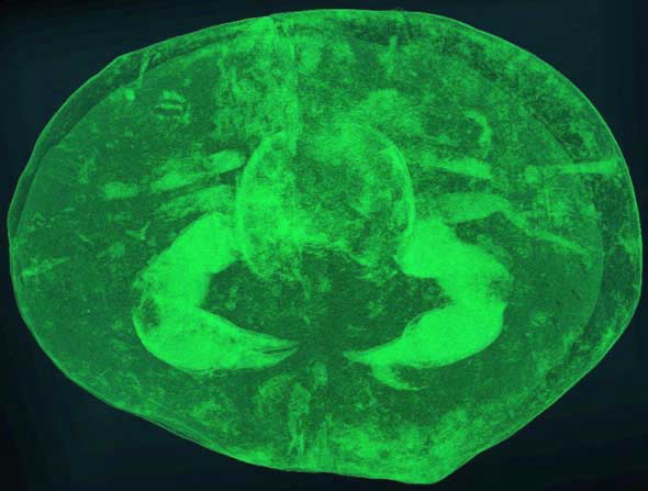
In this case, neutron-CT revealed the orientation of the crab, enabling careful preparation of the specimen out of the rock, but also that some of the legs were missing, which would make it unsuitable for museum display. The team then made another unusual observation.
We could see tunnels dug by worms that had ingested the innards of the crab and then tunnelled out again. We didn’t know then that this was the first time such processes had been captured with CT.”
Although a leg bone of an 18-metre long 8,500 kilogram dinosaur, Wintonotitan , proved to be too large and dense for neutron transmission, they had success with rock containing bones from another sample, a Cretaceous crocodylomorph, an ancestor or close relative to modern-day crocodiles.
“These creatures were similar to modern crocodiles, in that they were incredibly powerful and quick. They likely ate small dinosaurs along with any other available prey,” said Bevitt.
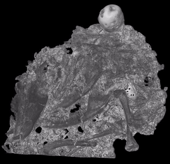
During early tests, a single rock sample was supplied for neutron CT. The researchers had assumed that sample contained only a single bone to be excavated. A three-dimensional CT reconstruction of the sample (depicted below) revealed that there were in fact many smaller bones contained in the rock —essentially an entire, articulated leg and foot.
Queensland has another important fossil site at Riversleigh, where there is a 15 million year old network of cave deposit with bones from extinct marsupials which walked and glided around a lush rainforest.
Researchers at the University of New South Wales were interested in doing quantitative bone measurements on an ancient, extinct bandicoot to better understand how bandicoots have evolved to their current form.
Important and delicate Riversleigh fossils are removed from limestone rock using dilute acid (similar in composition to vinegar), and special glues are used to preserve the 3D layout of those fossils. Although the glue used to preserve and protect the specimens was problematic for neutron radiography, the team were able to successfully use neutron tomography to image individual bone components for a full 3D reconstruction.
Further afield
Bevitt is excited by some interesting finds from Central Mongolia, where there is a Korea-Mongolia International Dinosaur Project that began in 2006.
He is working with the team of researchers to conduct CT scans on remarkably preserved dinosaur remains.
“Our understanding of dinosaurs has changed immensely over the past 10 years to the point that we are developing a picture of their rate of growth, lifespan, reproductive behaviour, colour and how they digested their food,” said Bevitt.
It is likely to be a combination of advanced techniques, neutron CT, synchrotron X-ray CT and Raman CT that provides the most valuable information, such as locating the presence of preserved soft tissue remains (proteins, but not DNA) determining the gender of individual dinosaur specimens and understanding how juvenile, and embryonic dinosaurs grew at such an incredible rate. “Alas, Jurassic Park is still not feasible,” said Bevitt.
“With these successes, we now do imaging of specimens from around the world—New Zealand, South Africa, China, the USA and Antarctica, covering the entire range of vertebrate evolution, and some of the very earliest invertebrate life forms,” said Bevitt.
 |
Other projects include the CT-scanning of fossilised eggs (see below right) and embryos of Lufengosaurus, one of the earliest known dinosaurs that lived in what is now southwestern China; imaging the fossilised brain and facial muscle structure of an early mammal-like reptile; and revealing a massive jaw bone and teeth of an new species of marine reptile found in East Timor that lived during the earliest age of dinosaurs, around 200 million years ago.
Published: 26/02/2016


